Improvement of early diagnostics, testing method “IgA t-TG deposits in tissue samples
University medical center Maribor – PP2 – UKC MB
New methods can detect changes at an earlier stage or in minor damage, which could be missed with basic histopathological examination.
Diagnosing celiac disease in majority of cases relies strongly on histopathological examination of intestinal mucosa. To obtain adequate tissue samples upper gastrointestinal endoscopy (EGDS) with multiple biopsies must be performed. Histopathological examination includes the description of the damage of intestinal villi, elongation of crypts, and increase in the number of intraepithelial lymphocytes. Newer methods are becoming available in some centers that enable detection of more subtle changes. These methods can detect changes at an earlier stage or in minor damage, which could be missed with basic histopathological examination. One of these methods is detection of deposits of celiac disease specific autoantibodies against tissue transglutaminase in intestinal mucosa.
We are therefore aiming to implement the method of detecting tissue deposits of tTG antibodies in our institution and in our country.
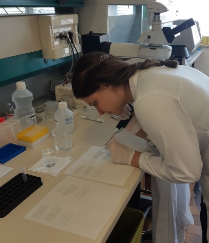
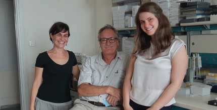
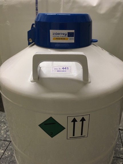
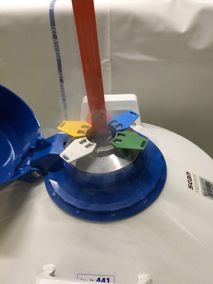
For that purpose, we purchased several containers in which we are able to store tissue samples at extremely low temperatures (liquid Nitrogen), which enable preservation of the samples for longer periods, as well as containers and freezers to enable transportation of the samples to local and distant labs. One of the members of our staff will in September learn the technique in a partner lab of IRCCS Burlo Garofolo in Trieste, and will later with the collaboration of personnel in Department of pathology and Department of Laboratory diagnostics of our institution, transfer the technique to Slovenia. This will improve the management of patients with mild or early changes who can be missed with the use of currently available diagnostic techniques.


